Stimuli and responses
What this chapter covers
- Describe the structure of the human nervous system and the pathway of impulses in voluntary and involuntary actions (1.1)
- Explain why the nervous system is important and measure reaction time (1.1)
- Describe the parts of the eye, ear, nose, tongue and skin and their functions (1.2)
- Explain the mechanisms of hearing and sight and the sensitivity of the skin and tongue to stimuli (1.2)
- Explain limitations of the senses, defects of sight and hearing and how they are corrected (1.2)
- Describe tropism and nastic movement in plants and how they help plants survive (1.3)
- Explain stereoscopic and monocular vision, stereophonic hearing and hearing ranges of animals (1.4)
1.1Human nervous system
Example: in badminton the moving shuttlecock is the stimulus. The eye detects it, an impulse goes to the brain, the brain judges its speed and direction, and impulses are sent to the hand and leg muscles, which respond by moving the body (1.1.1).
Structure of the nervous system
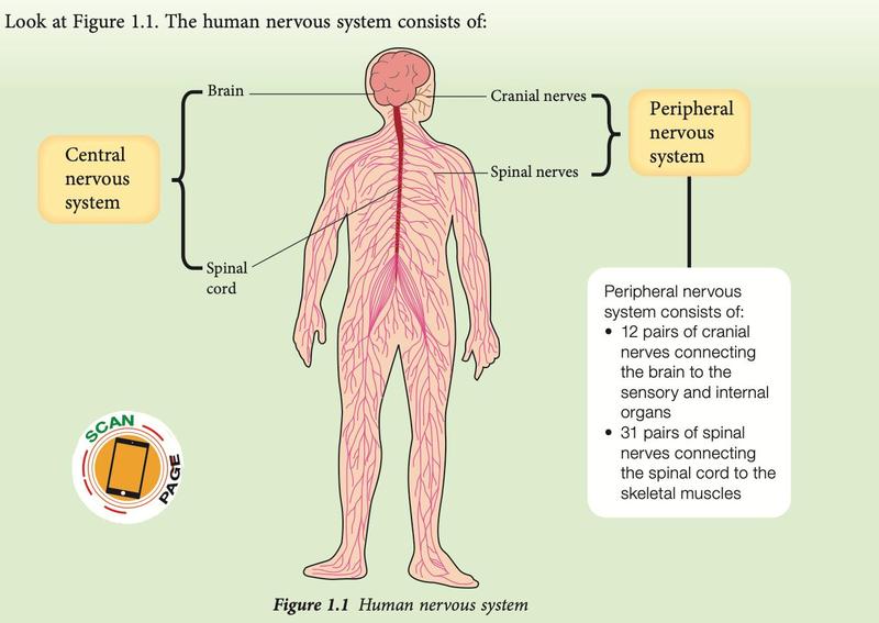
| Part | Made up of | Role |
|---|---|---|
| Central nervous system (CNS) | Brain and spinal cord | Interprets impulses and decides the response |
| Peripheral nervous system (PNS) | 12 pairs of cranial nerves (brain to sensory and internal organs) and 31 pairs of spinal nerves (spinal cord to skeletal muscles) | Carries impulses to and from the CNS |
Voluntary and involuntary actions (1.1.2)
| Voluntary action | Involuntary action | |
|---|---|---|
| Control | Conscious, under one's will; controlled by the brain | Occurs immediately, without conscious control or prior thought |
| Examples | Reading, writing, speaking, eating, walking, running | Medulla oblongata: heartbeat, breathing, peristalsis, secretion of saliva. Spinal cord (reflex): withdrawing a hand from a hot object, withdrawing a foot from a sharp object, sneezing when dust enters the nose |
Example: a phone rings (stimulus) → ear → brain → muscles of the hand touch the screen.
Reflex: stimulus → affector (receptor) → nerve → spinal cord → nerve → effector → response. The impulse does not need to reach the brain first, so the response is faster.
Reaction time (Activity 1.1)
Reaction time is the time between a stimulus and the response. In the falling-ruler activity, a partner releases a half-metre rule without warning and you catch it. The distance the ruler falls before you catch it measures your reaction time: a shorter distance means a faster reaction. Repeat several times and find the average distance. Different students have different reaction times, and one person's reaction time also changes from one attempt to the next.
Importance of the nervous system (1.1.3): it controls and coordinates all organs and keeps the internal environment balanced. If nerves or the brain are damaged, a person may be paralysed temporarily, partially or completely and may need machines for breathing or heartbeat.
Show answerHide answer
1.2Stimuli and responses in humans
Humans detect stimuli such as light, sound and chemicals with five sensory organs: eyes (sight), ears (hearing), nose (smell), tongue (taste) and skin (touch). The skin is the largest sensory organ.
The eye
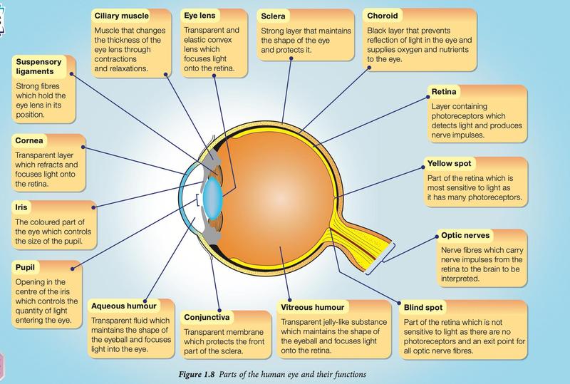
| Part | Function |
|---|---|
| Cornea | Transparent layer that refracts and focuses light onto the retina |
| Iris | Coloured part that controls the size of the pupil |
| Pupil | Opening in the centre of the iris that controls the quantity of light entering the eye |
| Aqueous and vitreous humours | Transparent fluid (aqueous) and jelly-like substance (vitreous) that keep the shape of the eyeball and help to focus light onto the retina |
| Conjunctiva | Transparent membrane that protects the front part of the sclera |
| Eye lens | Transparent, elastic convex lens that focuses light onto the retina |
| Ciliary muscle and suspensory ligaments | The muscle changes the thickness of the lens; the ligaments hold the lens in position |
| Retina | Contains photoreceptors that detect light and produce nerve impulses |
| Yellow spot | Part of the retina most sensitive to light (many photoreceptors) |
| Blind spot | No photoreceptors; the exit point of the optic nerve fibres |
| Optic nerve | Carries impulses from the retina to the brain |
| Sclera / choroid | Sclera: strong layer that keeps the eye's shape. Choroid: black layer that prevents reflection of light and supplies oxygen and nutrients |
- Rod cells: sensitive to different light intensities, including faint light, but not to colour.
- Cone cells: sensitive to colour in bright light; three types, for red, green and blue light.
- Pupil and light (Activity 1.3): in bright light the pupil becomes smaller, which protects the retina from excessive light. In dim light the pupil becomes larger so that more light enters. This is an involuntary response.
The ear
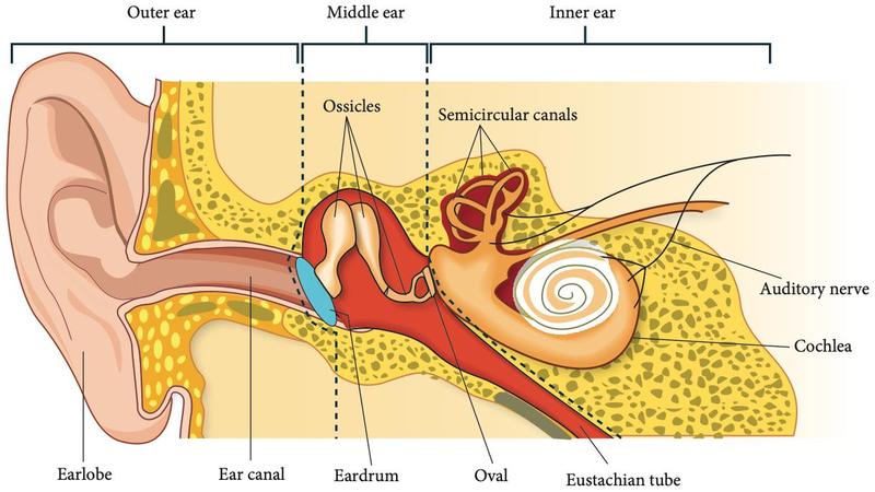
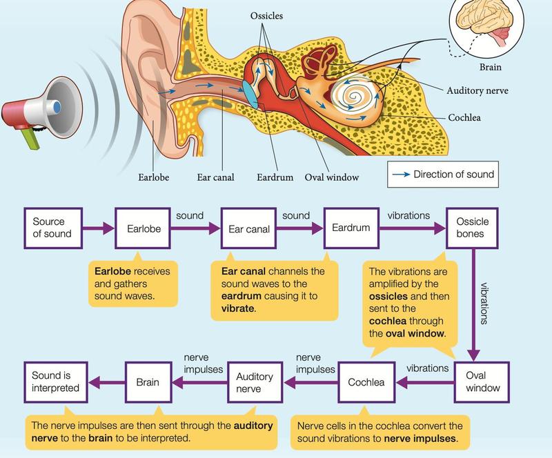
| Region | Part | Function |
|---|---|---|
| Outer ear | Earlobe | Collects sound waves and directs them into the ear canal |
| Ear canal | Directs sound waves to the eardrum | |
| Middle ear | Eardrum | Vibrates with the sound waves and passes the vibrations to the ossicles |
| Ossicles (3 small bones) | Amplify the vibrations and pass them to the oval window | |
| Oval window | Collects the vibrations from the ossicles and transfers them to the cochlea | |
| Eustachian tube | Balances the air pressure on both sides of the eardrum | |
| Inner ear | Cochlea | Converts sound vibrations to nerve impulses |
| Semicircular canals | Detect the position of the head and help balance the body | |
| Auditory nerve | Sends impulses from the cochlea to the brain |
Nose, tongue and skin
- Nose: about 10 million sensory cells for smell at the roof of the nasal cavity, covered with mucus. Chemicals in the air dissolve in the mucus and stimulate the cells; impulses go to the brain to be interpreted as a smell.
- Tongue: papillae on the surface carry hundreds of taste buds; each taste bud has 10 to 50 taste receptors. Chemicals dissolved in saliva diffuse into the taste buds through the pores and stimulate the receptors. Five basic tastes: sweet, salty, sour, bitter and umami.
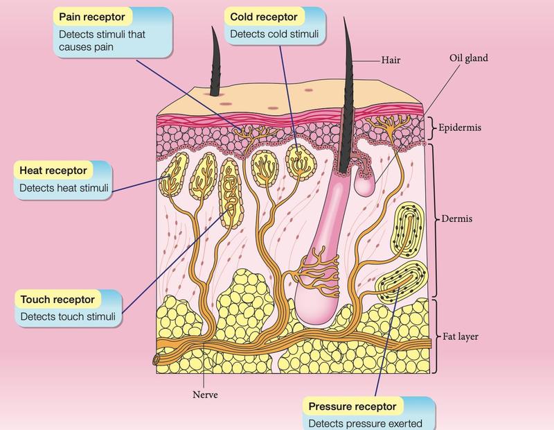
- Skin: thin outer epidermis and inner dermis. Five types of receptors detect pain, cold, heat, touch and pressure. The tongue, nose and lips are also very sensitive to touch.
Mechanism of hearing (1.2.2)
Mechanism of sight
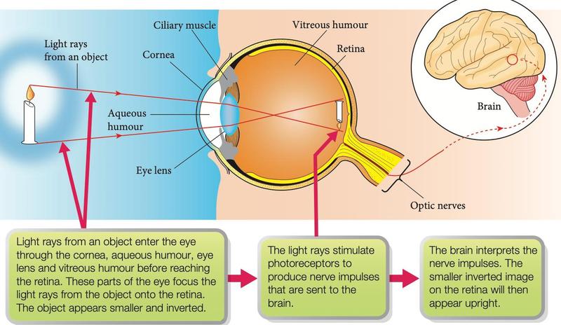
The image on the retina is smaller and inverted; the brain interprets it so that the object appears upright.
Sensitivity of the skin and the tongue (1.2.3)
- Skin sensitivity depends on the number of receptors and the thickness of the epidermis. The fingertip is very sensitive to touch: many touch receptors and a thin epidermis. The elbow, the sole of the foot and the back are less sensitive.
- In the toothpick activity, the part of the body where two toothpicks (0.5 cm apart) are felt as two points is the most sensitive.
- All areas of the tongue detect all five tastes, but the tip is more sensitive to sweet, the sides to sour and salty, the back to bitter and the centre to umami.
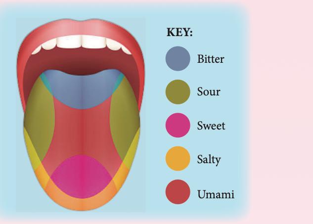
Taste and smell work together. With the nose pinched, a person finds it hard to identify the flavour of a drink. Smell therefore adds to the sense of taste, which is why food tastes less good when you have flu.
Limitations and defects of sight and hearing (1.2.4)
| Limitation or defect | Explanation |
|---|---|
| Very small or very distant objects | The eye cannot see tiny objects such as microorganisms or very distant objects such as the planet Jupiter; devices such as microscopes and binoculars overcome this |
| Optical illusion | An object seen differs from its actual state, because the brain cannot interpret it correctly when there are distractions around it |
| Blind spot | An image falling on the blind spot (no photoreceptors) cannot be seen; we do not notice because the same object cannot fall on the blind spots of both eyes at once |
| Human hearing range | Only 20 Hz to 20 000 Hz. The range narrows with age because the eardrum becomes less elastic |
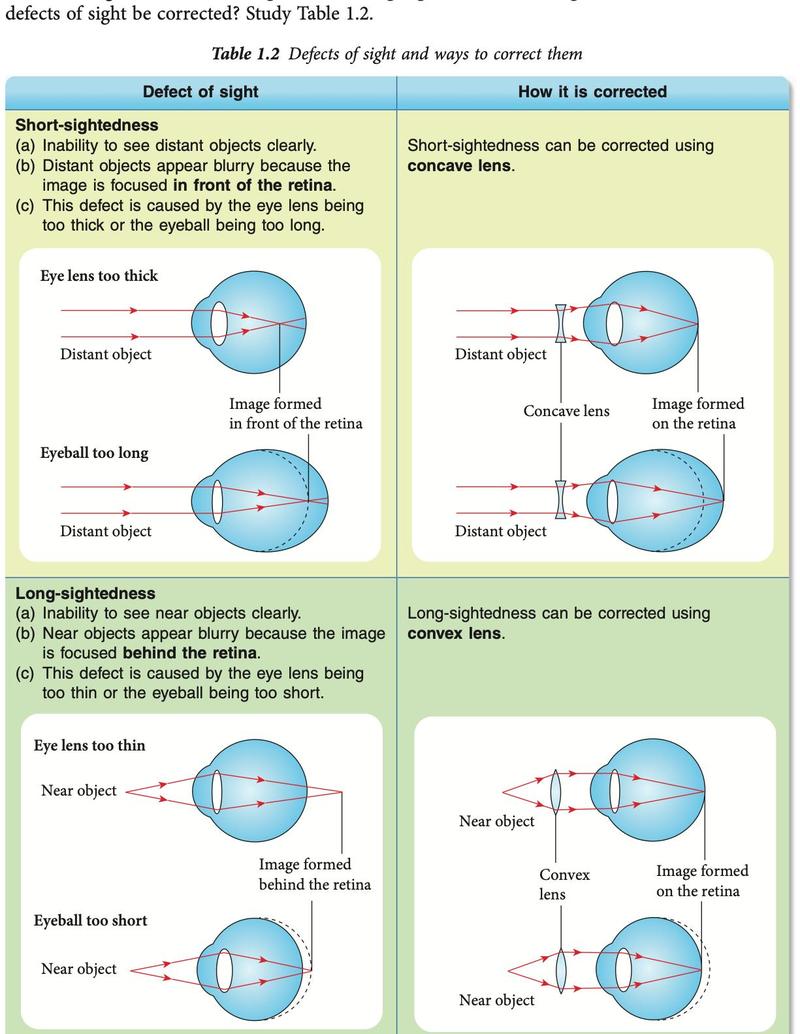
| Defect | Problem | Cause | Correction |
|---|---|---|---|
| Short-sightedness | Cannot see distant objects clearly; image forms in front of the retina | Eye lens too thick or eyeball too long | Concave lens |
| Long-sightedness | Cannot see near objects clearly; image forms behind the retina | Eye lens too thin or eyeball too short | Convex lens |
| Astigmatism | Part of an object is clearer than the rest | Uneven curvature of the cornea or eye lens | Cylindrical lens |
- Hearing defects come from infection, injury, ageing or loud sounds. Outer and middle ear damage (blocked canal, punctured eardrum, damaged ossicles) can be corrected by clearing, medicine or surgery. A damaged cochlea can be helped with a cochlear implant, but a damaged auditory nerve cannot be corrected.
- Devices that overcome limits: binoculars, microscopes, X-ray and ultrasound machines (sight); loudspeaker, stethoscope and hearing aids (hearing).
- The five senses are a gift. Unhealthy habits and high-risk jobs can damage them, so use protection such as safety goggles and ear protectors (1.2.5).
Show answerHide answer
1.3Stimuli and responses in plants
Plants detect light, water, gravity and touch. Their responses are tropism and nastic movement (1.3.1, 1.3.2).
| Tropism | Stimulus | Response | Importance |
|---|---|---|---|
| Phototropism | Light | Shoots: positive (grow towards light) | Shoots and leaves get sunlight for photosynthesis |
| Geotropism | Gravity | Roots: positive (grow down). Shoots: negative (grow up) | Roots grip and stabilise the plant; shoots reach sunlight |
| Hydrotropism | Water | Roots: positive (grow towards water) | Roots obtain water and mineral salts |
| Thigmotropism | Touch | Tendrils and twining stems: positive. Roots: negative (avoid obstacles) | Plant climbs to the light and gets support |
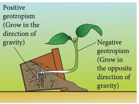
Gravity: seedlings arranged in different positions on moist cotton wool in a Petri dish, kept in a dark cupboard (so that light is not a second stimulus). Result: roots grow down, shoots grow up.
Water: one beaker has water below the wire gauze, the other has anhydrous calcium chloride (which absorbs water, so there is no water source). Roots grow towards the water.
| Tropism | Nastic movement |
|---|---|
| Direction of response depends on the direction of the stimulus | Direction does not depend on the direction of the stimulus |
| Slow | Rapid |
Similarity: both are responses of plants to stimuli.
Show answerHide answer
1.4Importance of responses to stimuli in animals
| Stereoscopic vision | Monocular vision | |
|---|---|---|
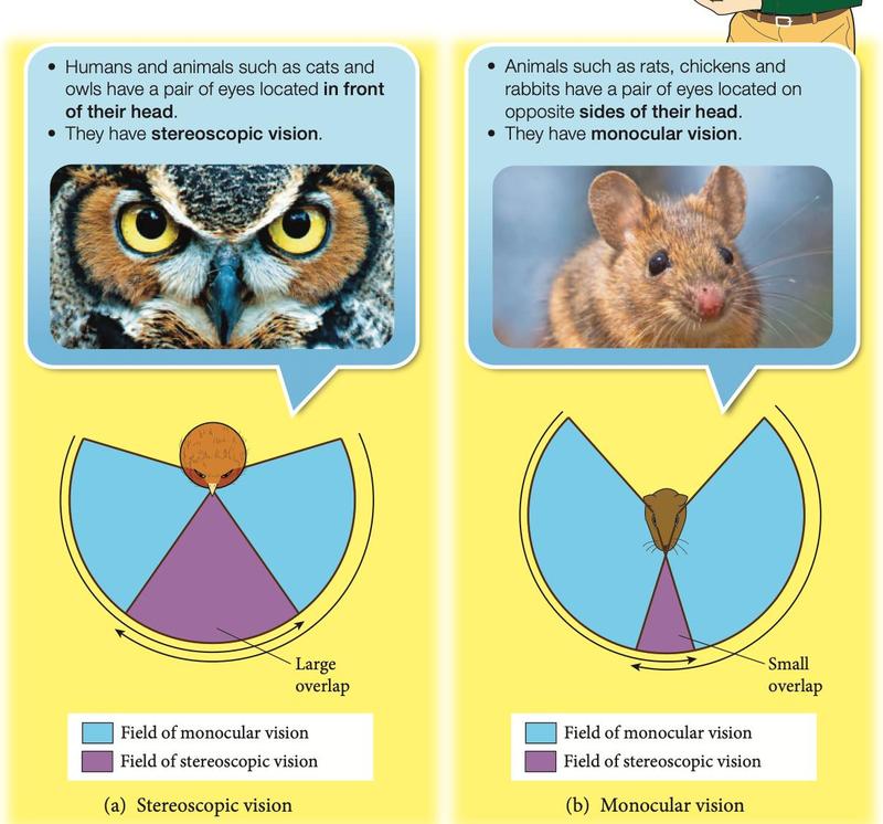
| Position of eyes | Both eyes in front of the head | Eyes at the sides of the head |
| Field of vision | Narrow; fields overlap a lot | Wide; little or no overlap |
| Image | Three-dimensional: distance, size and depth estimated accurately | Two-dimensional: distance and depth not estimated accurately |
| Advantage | Helps to hunt prey | Detects enemies from any direction |
| Examples | Humans, cats, owls; most predators | Rats, chickens, rabbits; most prey |
Stereophonic hearing
Hearing with both ears. The ear nearer to the source receives the sound earlier and louder. The brain detects the difference in time and loudness and works out the direction of the source. Predators use it to locate prey; prey use it to locate predators and escape.
Hearing frequencies of animals
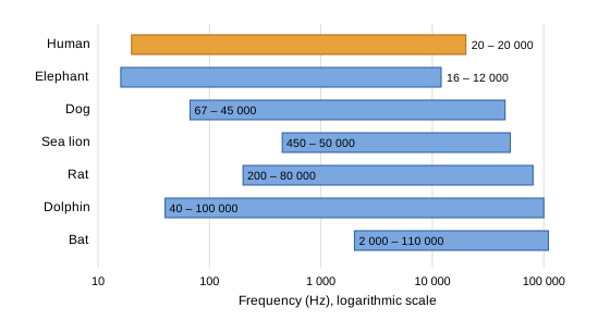
Bats hear the highest frequencies (up to 110 000 Hz). The elephant hears down to 16 Hz, which is below the human limit of 20 Hz. Responses to stimuli through special sensory organs ensure the survival of animals, for example the lateral line of fish, pheromones of insects and the electric field of an electric eel (1.4.2).
Show answerHide answer
Summary
| Remember | Key fact |
|---|---|
| Nervous system | CNS (brain, spinal cord) and PNS (cranial and spinal nerves) |
| Voluntary | Controlled by the brain; conscious |
| Involuntary | Medulla oblongata (heartbeat, breathing) or spinal cord (reflex) |
| Short- and long-sightedness | Image in front of / behind the retina; concave / convex lens |
| Human hearing range | 20 Hz to 20 000 Hz |
| Tropisms | Photo (light), geo (gravity), hydro (water), thigmo (touch) |
| Nastic movement | Rapid; independent of the direction of the stimulus |
| Vision in animals | Stereoscopic: predators, 3-D. Monocular: prey, wide field |