Cell Division
What this chapter covers
- Define karyokinesis, cytokinesis, haploid, diploid, chromatin and homologous chromosomes (6.1)
- Describe the cell cycle, the stages of mitosis and cytokinesis in animal and plant cells (6.2)
- Explain why mitosis is necessary for growth, healing, regeneration and asexual reproduction (6.2)
- Explain the meaning and necessity of meiosis and describe meiosis I and II (6.3)
- Compare and contrast mitosis and meiosis (6.3)
- Explain how abnormal mitosis causes tumours and cancer, and how abnormal meiosis causes Down syndrome (6.4)
6.1Cell division
Body cells grow, divide and die, so dead cells must be replaced. The cells of an organism are of two kinds.
| Feature | Somatic cell | Gamete |
|---|---|---|
| What it is | Any body cell except gametes | Reproductive cell (sperm, egg) |
| Made by | Mitosis | Meiosis |
| Chromosome number | Diploid, 2n (human 46) | Haploid, n (human 23) |
Show answerHide answer
6.2Cell cycle and mitosis
The cell cycle is the sequence of events of DNA replication and cell division that gives two daughter cells. It has interphase (the longest part) and M phase.
| Phase | What happens |
|---|---|
| G1 | Cell grows; organelles and proteins are made; chromosomes are chromatin |
| S | DNA is replicated; each chromosome becomes two sister chromatids joined at the centromere |
| G2 | Cell keeps growing and gathers energy for division |
| M | Mitosis (prophase, metaphase, anaphase, telophase) then cytokinesis |
| Stage | Key events |
|---|---|
| Prophase | Chromatin shortens and thickens into visible chromosomes (two sister chromatids); nuclear membrane and nucleolus disappear; centrioles move to opposite poles; spindle fibres form |
| Metaphase | Chromosomes line up in a single row on the equatorial plane, held by spindle fibres |
| Anaphase | Centromeres divide; spindle fibres shorten and pull sister chromatids to opposite poles |
| Telophase | Chromatids (now daughter chromosomes) reach the poles and uncoil; nucleoli and nuclear membranes reform; spindle fibres disappear |
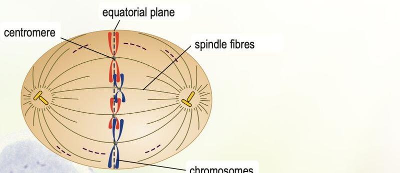
Textbook Figure 6.3, p. 103.
Cytokinesis in animal and plant cells
| Feature | Animal cell | Plant cell |
|---|---|---|
| Centrioles | Present | Absent (spindle fibres still form) |
| How the cytoplasm divides | Plasma membrane constricts in the middle (cleavage furrow) by a ring of microfilaments | Vesicles join to form a cell plate that grows outwards to the cell wall |
| New cell wall | None | Formed, strengthened with cellulose |
Why mitosis is necessary
- Embryo development and growth of organisms
- Healing of wounds: new cells replace dead or damaged ones
- Regeneration, for example a lizard regrowing a tail
- Asexual reproduction, for example budding in hydra
Applications of mitosis: in medicine, stem cell therapy uses bone marrow stem cells to treat damaged cartilage; in agriculture, plant tissue culture grows young plants from parent cells without fertilisation, so many identical, high quality plants are produced quickly.
Show answerHide answer
6.3Meiosis
Meiosis is necessary to form gametes, to keep the diploid number the same from one generation to the next, and to produce genetic variation.
| Stage | Key events |
|---|---|
| Prophase I | Homologous chromosomes pair (synapsis) to form a bivalent (tetrad); crossing over at chiasmata exchanges genetic material between non-identical chromatids |
| Metaphase I | Homologous pairs line up on the equatorial plane |
| Anaphase I | Homologous chromosomes separate and move to opposite poles; sister chromatids stay joined |
| Telophase I | Cytokinesis gives two haploid cells; DNA does not replicate before meiosis II |
| Meiosis II | Like mitosis: sister chromatids separate in anaphase II; four haploid cells result |
Mitosis compared with meiosis
| Feature | Mitosis | Meiosis |
|---|---|---|
| Where | Somatic cells | Reproductive organs |
| Divisions | One | Two (I and II) |
| Daughter cells | 2, diploid, identical to parent | 4, haploid, genetically different |
| Homologous pairing and crossing over | No | Yes (prophase I) |
| Metaphase | Single chromosomes in a row | Homologous pairs on the plane (metaphase I) |
6.4Cell division and human health
Abnormal mitosis. Uncontrolled division forms a tumour. A benign tumour is not dangerous and can be removed surgically. A malignant tumour is cancer: cells spread through lymph and blood vessels and destroy surrounding tissue. Causes include radiation (X-ray, gamma, UV), chemicals (tar, formaldehyde, benzene), genetic factors, bacteria and viruses.
Abnormal meiosis. If chromosomes fail to separate in anaphase I or II (nondisjunction), a gamete has 24 (or 22) chromosomes. Fertilisation with a normal gamete (23) gives a zygote of 47 chromosomes with three copies of chromosome 21: Down syndrome (trisomy 21). Such a person has 47 chromosomes and shows stunted growth, slanted eyes and a slightly protruding tongue.
Radiotherapy uses radiation to damage and kill dividing cancer cells, so it controls or stops the growth of the tumour.
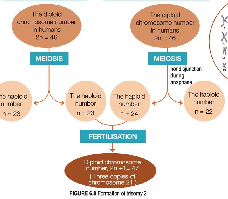
Textbook Figure 6.8, p. 109.