Identify respiratory structures in insects, fish, amphibians and humans (8.1.1)
Describe how respiratory structures are adapted for gaseous exchange, and compare them (8.1.2, 8.1.3)
Compare and contrast breathing mechanisms in insects, fish, frogs and humans (8.2.1)
Explain gaseous exchange between lungs and blood and between blood and tissues, and how oxygen and carbon dioxide are transported (8.3.1)
Describe the effects of asthma, chronic bronchitis and emphysema (COPD) on the respiratory system (8.4.1)
8.1Types of respiratory system
Respiratory structure (respiratory surface) is the surface where gaseous exchange takes place between the cells of an organism and its outer environment. Breathing is the repeated process of inhalation and exhalation.
A unicellular organism such as Amoeba needs no special structure: its total surface area to volume ratio (TSA/V) is large, so gases diffuse across the cell membrane. The bigger an organism is, the smaller its TSA/V, because volume grows faster than surface area. Large, complex organisms therefore need a special respiratory structure.
Adaptations of respiratory structures in large organisms
Large TSA/V for efficient exchange of gases.
Thin, one cell thick, so gases diffuse a short distance.
Always moist, so that gases can dissolve.
Network of blood capillaries (except insects) to carry gases quickly.
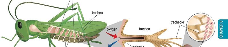
Insects: the tracheal system
The tracheal system in a grasshopper. (Textbook Figure 8.1, p. 129)
Air enters through spiracles (small pores in the thorax and abdomen) into the trachea, which branches into fine tracheoles.
The tracheole is the respiratory surface: very many, with thin, moist walls, so oxygen diffuses straight into body cells and carbon dioxide diffuses out.
Some insects have air sacs, filled with air to speed up gas delivery during active movement.
Air goes directly to the cells, so insects need no blood to carry respiratory gases.
Fish: gills
Gills consist of rows of filaments held by a gill arch. Each filament has many thin, flat lamellae (singular: lamella). Many filaments and lamellae give a large total surface area; the thin lamella membrane has many blood capillaries to absorb oxygen and carry away carbon dioxide.
A very large number of alveoli give a large total surface area.
The wall is one cell thick and always moist, so O2 and CO2 dissolve and diffuse through easily.
Each alveolus is surrounded by a dense network of blood capillaries.
Comparison
Feature
Insects
Fish
Frogs
Humans
Respiratory structure
Tracheole
Filament and lamella
Skin and lungs
Alveolus
Large TSA/V by
Many tracheoles
Many filaments and lamellae
Folded lung surface; whole skin
Many alveoli
Blood capillaries
None
Yes
Yes
Yes
Insects do not use blood to carry oxygen. Gases go through the tracheal tubes directly to the cells.
A fish is caught in a net and its gills are torn. Predict what happens and explain.
Show answerHide answer
The total surface area of the gills falls, so less oxygen diffuses into the blood and the fish may suffocate.
8.2Mechanisms of breathing
Insects
Inhalation: abdominal muscles relax, pressure in the trachea falls, air enters through the spiracles.
Exhalation: abdominal muscles contract, pressure in the trachea rises, air is forced out through the spiracles.
Fish
Inhalation (mouth opens)
Exhalation (mouth closes)
Buccal cavity floor
Lowered
Raised
Opercular cavity / operculum opening
Enlarged / closed
Smaller / opened
Pressure in buccal cavity
Lower than outside
Higher than outside
Water flow
Enters mouth
Passes over the lamellae, then leaves through the operculum opening
Frogs (active state)
Inhalation: mouth and glottis closed, floor of the buccopharyngeal cavity lowered, air drawn in through the nostrils. Then the glottis opens, nostrils close, the floor is raised and air is pushed into the lungs.
Exhalation: lungs contract (abdominal pressure and elastic lungs); air leaves through the nostrils, some mixing with air in the buccopharyngeal cavity.
Humans
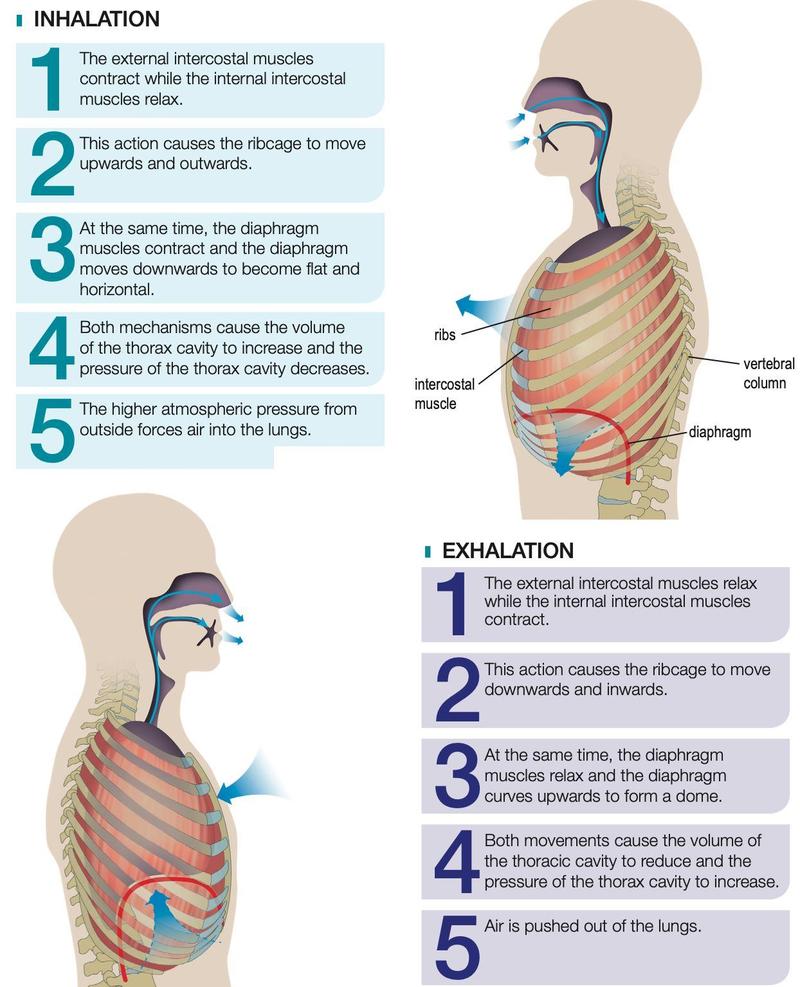
Inhalation and exhalation in humans. (Textbook Figure 8.5, p. 134)
Step
Inhalation
Exhalation
Intercostal muscles
External contract, internal relax
External relax, internal contract
Ribcage
Up and out
Down and in
Diaphragm
Contracts, flattens
Relaxes, dome shape
Thoracic volume
Increases
Decreases
Thoracic pressure
Decreases; air pushed in by higher atmospheric pressure
Increases; air pushed out
Every breathing mechanism changes volume and pressure of the respiratory cavity: low pressure draws air or water in, high pressure pushes it out.
State two differences between breathing in fish and in humans.
Show answerHide answer
Fish use mouth and operculum; humans use nostrils. Fish use the muscular floor of the buccal cavity and operculum; humans use the diaphragm, ribcage and intercostal muscles. (Any two.)
8.3Gaseous exchange in humans
Gases diffuse down a partial pressure gradient: from where their partial pressure is higher to where it is lower.
At sea level atmospheric pressure is 760 mm Hg and air is 21% oxygen, so the partial pressure of oxygen is 0.21 × 760 = about 160 mm Hg.
Lungs and blood
Blood arriving in the lung capillaries (pulmonary artery) has low oxygen and high carbon dioxide partial pressure.
O2 diffuses from the alveolus into the capillary and combines with haemoglobin in erythrocytes to form oxyhaemoglobin.
CO2 diffuses from the capillary into the alveolus and is breathed out.
Blood leaving through the pulmonary vein is rich in oxygen.
Blood and tissues
Cells use oxygen and release CO2 in cellular respiration, so oxygen is low and CO2 high in the cells.
Oxyhaemoglobin breaks down; O2 diffuses from the tissue capillaries into the cells.
CO2 diffuses from the cells into the tissue capillaries and is carried to the lungs.
How carbon dioxide is carried
Form
Share
Bicarbonate ion (HCO3-) in plasma
70%
Carbaminohaemoglobin
23%
Dissolved, as carbonic acid (H2CO3)
7%
At the tissues: CO2 + H2O forms H2CO3 in the erythrocyte (catalysed by carbonic anhydrase); H2CO3 breaks into HCO3- and H+; HCO3- diffuses into plasma.
At the lungs: HCO3- re-enters the erythrocyte, combines with H+ to form H2CO3, which breaks into CO2 and water; CO2 diffuses into the alveolus and is exhaled.
Oxygen travels mainly as oxyhaemoglobin; carbon dioxide travels mainly as bicarbonate ions, not as oxyhaemoglobin.
Why does oxygen diffuse from the alveolus into the blood?
Show answerHide answer
The partial pressure of oxygen in the alveolus is higher than in the lung capillary blood, so it diffuses down the gradient.
8.4Health issues of the respiratory system
Chronic Obstructive Pulmonary Disease (COPD) includes asthma, chronic bronchitis and emphysema.
Disease
What happens
Effect
Asthma
Bronchiole walls swell and thicken; the air passage narrows
Difficulty breathing, breathlessness
Chronic bronchitis
Bronchioles inflamed, swollen and blocked; much mucus; damaged cilia cannot clear it
Reduced airflow, continuous coughing
Emphysema
Alveolus walls damaged; alveoli lose elasticity and enlarge
Total surface area falls, gaseous exchange less efficient
Explain why gaseous exchange is less efficient in an emphysema patient.
Show answerHide answer
The damaged alveoli lose elasticity and enlarge, so the total surface area for exchange decreases and less oxygen diffuses into the blood.