Transport in Humans and Animals
What this chapter covers
- Justify why large multicellular organisms need a transport system, and compare open and closed circulatory systems in insects, fish, amphibians and humans (10.1)
- Describe the heart, the blood vessels and the composition of blood, and compare arteries, capillaries and veins (10.2)
- Explain the heartbeat mechanism and the forces that move blood around the body (10.3)
- Describe blood clotting and the health issues thrombosis, embolism and haemophilia (10.4)
- Explain the ABO blood groups, the Rhesus factor and its effect in pregnancy (10.5)
- Describe cardiovascular diseases and how to keep the circulatory system healthy (10.6)
- Explain how tissue fluid and lymph form, the parts of the lymphatic system and why it is needed (10.7)
- Describe health issues related to the lymphatic system such as oedema and lymphatic filariasis (10.8)
10.1Types of circulatory system
A unicellular organism such as Amoeba has a large TSA/V, so diffusion across its surface is enough. In vertebrates the transport system is the blood circulatory system.
| Feature | Insects | Fish | Amphibians | Humans |
|---|---|---|---|---|
| Type | Open | Closed | Closed | Closed |
| Circulations per cycle | Not applicable (open system) | Single | Double (incomplete) | Double (complete) |
| Heart | Many segments with openings (ostia) | 2 chambers: 1 atrium, 1 ventricle | 3 chambers: 2 atria, 1 ventricle | 4 chambers: 2 atria, 2 ventricles |
| Oxygenated and deoxygenated blood | Haemolymph bathes the cells directly in the haemocoel | Not applicable (one circuit: gills, then body) | Partly mixed in the single ventricle | Completely separated by the septum |
- Open system: haemolymph leaves the heart through open-ended vessels, flows into the body cavity (haemocoel) and bathes the cells directly. It returns to the heart through the ostia when the heart relaxes.
- Closed system: blood stays inside continuous vessels. Exchange with cells takes place across the thin walls of blood capillaries.
- Fish (single): ventricle, then gill capillaries, then body (systemic) capillaries, then atrium. Blood passes through the heart once per cycle, so its pressure drops after the gills.
- Amphibians (incomplete double): pulmocutaneous circulation (lungs and skin) and systemic circulation. One ventricle lets some mixing happen.
- Humans (complete double): pulmonary circulation (heart, lungs, heart) and systemic circulation (heart, body, heart). The septum prevents mixing.
Show answerHide answer
10.2Circulatory system of humans
Three components: blood (the transport medium), the heart (a muscular pump) and blood vessels (arteries, capillaries, veins).
Structure of the heart
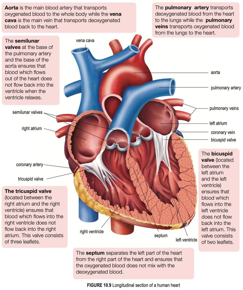
Textbook Figure 10.9, p. 174.
| Part | Function |
|---|---|
| Atria | Receive blood returning to the heart |
| Ventricles | Pump blood out; thicker walls than atria |
| Left ventricle | Thickest wall: pumps blood at high pressure to the whole body through the aorta |
| Septum | Separates left and right sides so the two kinds of blood do not mix |
| Tricuspid valve (3 flaps) | Between right atrium and right ventricle; stops backflow |
| Bicuspid valve (2 flaps) | Between left atrium and left ventricle; stops backflow |
| Semilunar valves | At the base of the aorta and pulmonary artery; stop blood returning to the ventricles |
| Aorta / vena cava | Main artery to the body / main vein returning deoxygenated blood |
| Pulmonary artery / pulmonary vein | Deoxygenated blood to the lungs / oxygenated blood to the left atrium |
| Coronary artery / vein | Supply oxygenated blood to the heart muscle / remove deoxygenated blood from it |
Composition of blood
Blood is about 55% plasma and 45% cells. Plasma is about 90% water and carries dissolved nutrients, urea, respiratory gases, hormones, enzymes and plasma proteins (fibrinogen for clotting, albumin for osmotic pressure, globulin as antibodies).
| Cell | Features | Function |
|---|---|---|
| Erythrocyte (red blood cell) | Biconcave disc, no nucleus when mature, contains haemoglobin, lives about 120 days | Carries oxygen as oxyhaemoglobin; biconcave shape gives large TSA/V and room for more haemoglobin |
| Leucocyte (white blood cell) | Has a nucleus, no haemoglobin, irregular shape, can leave capillaries | Defence: phagocytosis (neutrophils, monocytes) and antibody production (lymphocytes) |
| Platelet | Cell fragments from bone marrow, short life | Blood clotting |
Blood vessels
| Feature | Artery | Capillary | Vein |
|---|---|---|---|
| Wall | Thick, muscular, elastic | One cell thick | Thin, less muscular |
| Lumen | Small | Very tiny | Large |
| Valves | None (except semilunar valves at the heart) | None | Present |
| Blood pressure | High | Low | Very low |
| Direction | From the heart | Artery to vein, exchange with cells | To the heart |
Show answerHide answer
10.3Mechanism of heartbeat
- The SA node generates electrical impulses.
- The impulses spread through both atria, which contract together and push blood into the ventricles.
- The impulses reach the atrioventricular (AV) node, then pass along the bundle of His and the Purkinje fibres to the apex.
- The ventricle walls contract from the apex upwards and pump blood into the aorta and pulmonary artery.
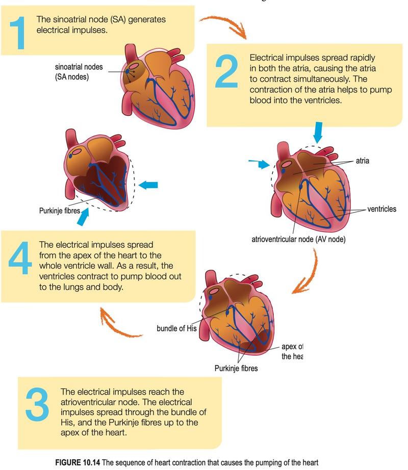
Textbook Figure 10.14, p. 181.
The "lub" sound is the tricuspid and bicuspid valves closing; the "dub" sound is the semilunar valves closing.
The pumping of the heart alone cannot push blood back through the veins against gravity. Blood returns with the help of valves, the contraction of skeletal muscles around the veins and the contraction of smooth muscle in the walls of venules and veins. Contracting skeletal muscles squeeze the vein, so blood is pushed towards the heart, and the valves close to stop backflow.
Show answerHide answer
10.4Mechanism of blood clotting
Clotting stops blood loss, keeps blood pressure normal and stops microorganisms entering the wound.
- Platelets, damaged cells and clotting factors form thrombokinase.
- With calcium ions and vitamin K, thrombokinase converts inactive prothrombin to thrombin.
- Thrombin (an enzyme) converts soluble fibrinogen to insoluble fibrin threads.
- Fibrin forms a mesh that traps erythrocytes and seals the wound.
| Condition | What happens |
|---|---|
| Haemophilia | Hereditary; some clotting factors are missing, so even small wounds bleed heavily |
| Thrombosis | A clot (thrombus) forms inside a vessel, from damaged vessel walls or slow blood flow |
| Embolism | A clot carried by the blood (embolus) blocks a small vessel and stops blood flow |
Show answerHide answer
10.5Blood groups of humans
| Group | Antigen on red cells | Antibody in serum | Can donate to | Can receive from |
|---|---|---|---|---|
| A | A | Anti-B | A, AB | A, O |
| B | B | Anti-A | B, AB | B, O |
| AB | A and B | None | AB only | A, B, AB, O |
| O | None | Anti-A and anti-B | A, B, AB, O | O only |
If the donor and recipient are not compatible, the recipient's antibodies make the donor's red blood cells clump together (agglutination).
Rhesus factor
People with antigen D on their red cells are Rh-positive; those without are Rh-negative. An Rh-negative person exposed to Rh-positive blood makes anti-D antibodies, and a second dose of Rh-positive blood causes agglutination.
In pregnancy, an Rh-negative mother with an Rh-positive foetus may make anti-D antibodies near birth. These are too few to harm the first baby, but in a second Rh-positive pregnancy they cross the placenta and destroy the foetus's red cells (erythroblastosis fetalis). Treatment is anti-Rhesus globulin given to the mother after the first pregnancy.
Show answerHide answer
10.6Health issues of the circulatory system
Keep the circulatory system healthy with a low-fat balanced diet and regular exercise, and by not smoking or drinking alcohol.
| Disease | Explanation |
|---|---|
| Atherosclerosis | Plaque (cholesterol, lipids, dead muscle cells, platelets) builds up on artery walls and narrows the lumen |
| Arteriosclerosis | Calcium deposits on the plaque make the artery hard and non-elastic |
| Hypertension | High blood pressure from narrowed arteries; can rupture fine arteries |
| Stroke | Blood supply to part of the brain is blocked by a clot, or a vessel in the brain bursts |
| Angina | Narrowed coronary artery gives too little oxygen to heart muscle; chest pain |
| Myocardial infarction | Coronary artery completely blocked; part of the heart muscle dies (heart attack) |
Show answerHide answer
10.7Human lymphatic system
- At the arteriole end of a capillary the blood pressure is high, so plasma leaks into the spaces between cells and becomes tissue fluid.
- Tissue fluid has no erythrocytes, platelets or plasma proteins (too large to leave). Oxygen and nutrients diffuse from it to cells; waste and carbon dioxide diffuse the other way.
- At the venule end, pressure is lower and the plasma is hypertonic, so most (about 85%) of the fluid returns to the blood.
- The remaining fluid (about 15%) enters the blind-ended lymphatic capillaries and is called lymph.
| Compared with | Lymph |
|---|---|
| Tissue fluid | More fat and fat-soluble substances, more lymphocytes |
| Blood | No plasma protein, erythrocytes or platelets |
Lymph flows through lymphatic vessels (with valves and lymph nodes) into the thoracic duct and the right lymphatic duct. These empty into the left and right subclavian veins. There is no pump: lymph is moved by skeletal muscle contraction, heartbeat pulses, peristalsis and breathing movements. Lymphatic organs include lymph nodes, spleen, thymus, bone marrow, tonsils and appendix.
Why the lymphatic system is needed
- Returns excess tissue fluid to the blood, keeping blood volume and pressure normal.
- Transports fats and fat-soluble substances (lacteals in the villi).
- Body defence: lymph nodes make and store lymphocytes.
Show answerHide answer
10.8Health issues of the lymphatic system
Oedema is swelling caused when tissue fluid is not returned to the blood. Causes include a blocked lymphatic vessel, a low level of albumin in plasma, pregnancy, and long periods of being bedridden (little muscle movement). Lymphatic filariasis is caused by the parasitic worm Brugia that blocks lymphatic vessels; it is spread by mosquito bites and swells the legs.