Transportation
What this chapter covers
- Describe why simple organisms need no transport system while complex organisms do, and justify the importance of transport (3.1)
- Compare the blood circulatory systems of vertebrates and explain the structure and functions of the human heart and blood vessels (3.2)
- Explain the double circulation, heartbeat, blood pressure and the factors that affect the pulse rate (3.2)
- Describe the components of human blood, the blood groups and compatible blood for transfusion, and the importance of blood donation (3.3)
- Explain transpiration and exudation, and investigate the factors that affect the rate of transpiration (3.4)
- Differentiate the structures and functions of xylem and phloem in vascular bundles (3.4)
- Compare the blood circulatory system in animals with the transport system in plants (3.5)
3.1Transport system in organisms
Every cell needs oxygen and nutrients for cellular respiration to release energy. The carbon dioxide and other wastes that cells make must be removed.
| Simple organisms | Complex organisms | |
|---|---|---|
| Examples | Amoeba, Euglena, Paramecium (unicellular) | Humans, vertebrates, multicellular plants |
| Transport system | None. Oxygen and nutrients diffuse in directly through the cell membrane; carbon dioxide diffuses out | Specialised system: blood circulatory system (animals), xylem and phloem (plants) |
| Why | The cell is small, so diffusion is fast enough | Large volume, so diffusion alone is too slow and does not reach all cells |
Importance of a transport system
- Carries oxygen and nutrients to all cells for respiration, so cells release energy for living processes.
- Carries mineral salts, water and the products of photosynthesis to plant cells.
- Eliminates toxic waste products from the cells. If wastes build up, they poison the cells and the organism can die.
Show answerHide answer
3.2Blood circulatory system
In vertebrates
All vertebrates have a blood circulatory system: blood flows in blood vessels through a heart that pumps it to the whole body and back. The number of heart chambers and the number of times blood passes through the heart in one cycle differ.
| Group | Heart chambers | Blood through the heart in one cycle |
|---|---|---|
| Fish | 1 atrium, 1 ventricle | Once (single circulation); blood goes heart, gills, body, heart |
| Amphibians | 2 atria, 1 ventricle | Twice (double circulation); oxygenated and deoxygenated blood mix in the ventricle |
| Reptiles | 2 atria, 1 ventricle | Twice (double circulation) |
| Mammals and birds | 2 atria, 2 ventricles | Twice (double circulation); no mixing of oxygenated and deoxygenated blood |
Based on textbook Figures 3.3 and 3.4, pp. 83–84.
The human heart
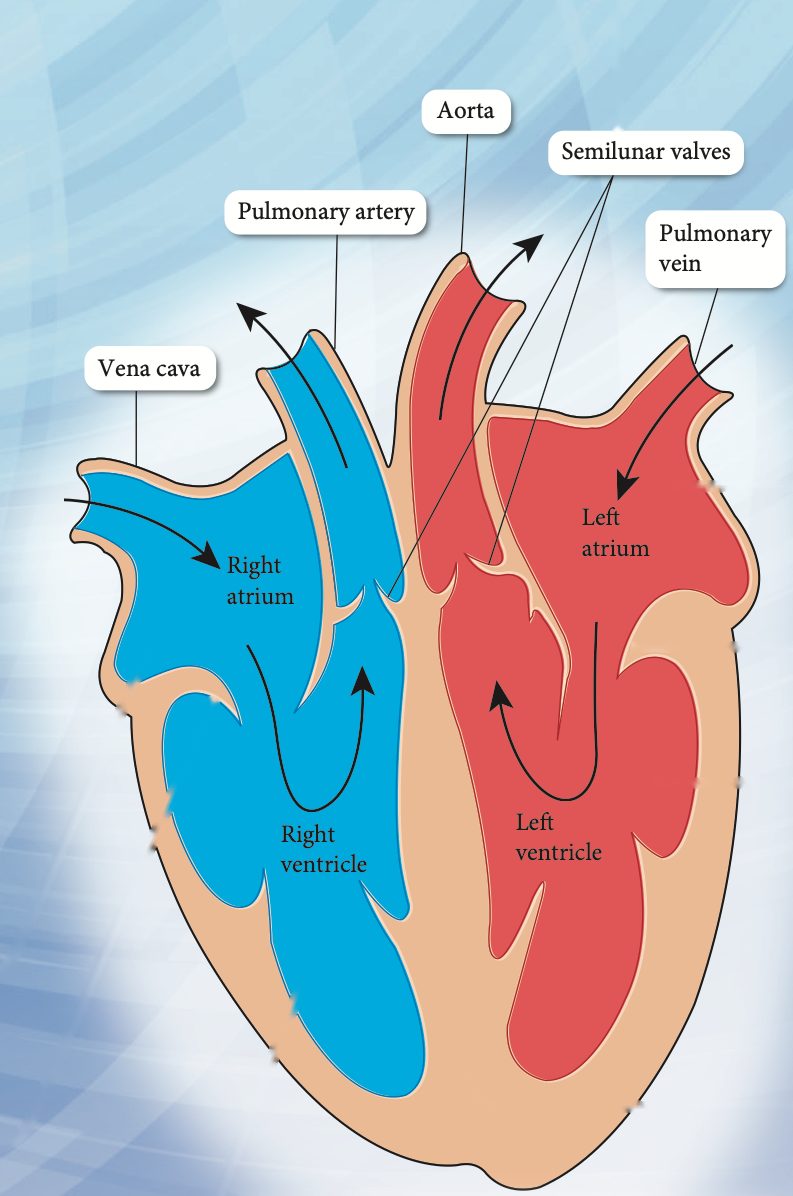
The heart has four chambers: two thin-walled atria that receive blood and two ventricles that pump it out. The septum separates the left side from the right side.
| Part | Function |
|---|---|
| Right atrium | Receives deoxygenated blood from the body (not the lungs) through the superior and inferior vena cava; contracts to push it into the right ventricle |
| Tricuspid valve | One-way flow from right atrium to right ventricle |
| Right ventricle | Pumps deoxygenated blood out through the pulmonary artery to the lungs |
| Left atrium | Receives oxygenated blood from the lungs through the pulmonary vein; contracts to push it into the left ventricle |
| Bicuspid valve | One-way flow from left atrium to left ventricle |
| Left ventricle | Thickest wall. Pumps oxygenated blood out through the aorta to all parts of the body except the lungs |
| Semilunar valves | At the pulmonary artery and aorta; stop blood flowing back into the ventricles |
| Septum | Muscular wall that stops oxygenated and deoxygenated blood mixing |
Show answerHide answer
Blood vessels
| Artery | Capillary | Vein | |
|---|---|---|---|
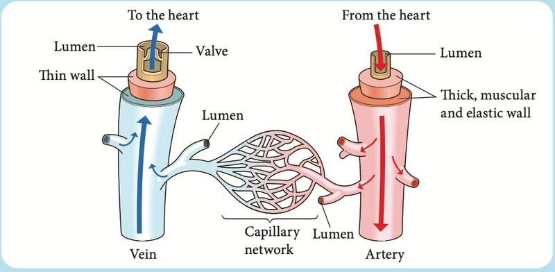
| Wall | Thick, muscular, lots of elastic tissue (withstands high pressure) | One cell thick; no muscle or elastic tissue | Thin, less muscular, less elastic |
| Lumen | Small | Smallest | Large |
| Valves | No | No | Yes |
| Function | Carries blood away from the heart (oxygenated, except the pulmonary artery) | Exchange of gases, food and wastes between blood and cells by diffusion | Carries blood back to the heart (deoxygenated, except the pulmonary vein) |
| Blood flow | Rapid, high pressure; pulse detected | Slow, falling pressure; no pulse | Slow, low pressure; no pulse |
The "double" circulation
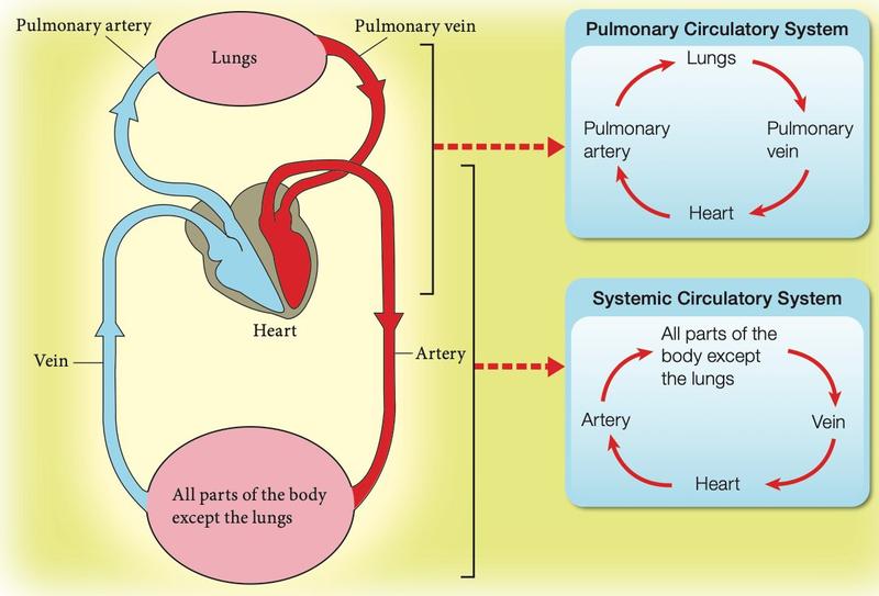
Humans and other mammals have two circuits that both pass through the heart.
| Circuit | Route | Purpose |
|---|---|---|
| Pulmonary | Heart, pulmonary artery, lungs, pulmonary vein, heart | Blood picks up oxygen and gives up carbon dioxide in the lungs |
| Systemic | Heart, aorta and arteries, all parts of the body except the lungs, veins, heart | Delivers oxygen and nutrients to cells and collects wastes |
Heartbeat and blood pressure
| Systole | Diastole | |
|---|---|---|
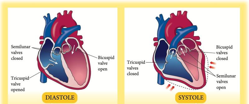
| Ventricles | Contract | Relax |
| Valves | Tricuspid and bicuspid close; semilunar open | Semilunar close; tricuspid and bicuspid open |
| Sound | "lub" (atrioventricular valves close) | "dub" (semilunar valves close) |
| Pressure reading | Systolic: blood flowing out of the heart (higher) | Diastolic: blood flowing into and filling the heart (lower) |
Blood pressure is measured with a sphygmomanometer. A youth normally has 120/75 mm Hg (systolic/diastolic). The pulse is produced by the contraction and relaxation of the muscular wall of an artery.
Factors that affect the pulse rate (Experiment 3.1)
- Physical activity — the more vigorous the activity, the higher the pulse rate. Muscles need more oxygen and must release more carbon dioxide, so the heart beats faster.
- Gender — adult males average 70 to 72 beats per minute and adult females 78 to 82, because a female heart is usually smaller and pumps less blood per beat.
- Age — the average maximum pulse rate falls as age rises (200 per minute at 20 years, 150 at 70).
- Body health — an unhealthy person may have a pulse rate that is too high or too low; both can be life-threatening.
Pulse rate per minute = 12 × 6 = 72 beats per minute (there are six 10-second periods in a minute).
Keeping the heart healthy
Heart disease has been one of the main causes of death in Malaysia since 2005 and is closely linked to diet and lifestyle. A healthy heart keeps the body supplied with oxygen and nutrients. Eat a balanced diet low in fat, exercise regularly and avoid smoking.
Show answerHide answer
3.3Human blood
Components of blood
Blood is a mixture. When spun in a centrifuge it separates into a yellow liquid (plasma) above a red layer (red blood cells).
| Component | Share of blood | Notes |
|---|---|---|
| Blood plasma | about 55% | About 90% water and 10% dissolved substances: nutrients, carbon dioxide, enzymes, hormones, wastes. The largest component |
| Red blood cells | about 45% | Carry oxygen |
| White blood cells and platelets | less than 1% | Body defence; platelets help blood to clot |
Blood groups
The four groups A, B, AB and O depend on the antigens on the red blood cells. The plasma carries antibodies. An antibody attacks the matching antigen and makes the blood coagulate (clump), which can kill.
| Blood group | Antigens on red blood cells | Antibodies in plasma |
|---|---|---|
| A | A | Anti-B |
| B | B | Anti-A |
| AB | A and B | None |
| O | None | Anti-A and Anti-B |
Textbook Table 3.3, p. 98.
Which blood can be given to whom
| Donor | Recipient A | Recipient B | Recipient AB | Recipient O |
|---|---|---|---|---|
| A | Yes | No | Yes | No |
| B | No | Yes | Yes | No |
| AB | No | No | Yes | No |
| O | Yes | Yes | Yes | Yes |
Textbook Table 3.4, p. 99.
Blood donation
- Blood is needed for surgery, accident victims and patients with leukaemia, haemophilia and other illnesses. Donated blood is stored in a blood bank.
- A donor must be healthy, aged 18 to 60 years and have a body mass of more than 45 kg. Up to 0.5 litres can be given at a time.
- After donating, the bone marrow makes new red blood cells.
Show answerHide answer
3.4Transport system in plants
Transpiration
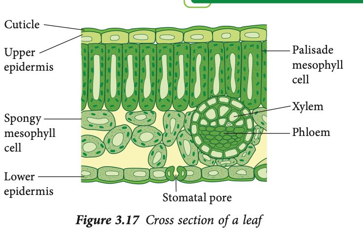
- The roots absorb water by osmosis, and the water moves up the plant to the leaves, where most of it is lost by transpiration.
- The waxy cuticle covers the epidermis and reduces water loss.
- Most water vapour leaves through the stomatal pores, which are surrounded by guard cells. The stoma is usually open during the day, when photosynthesis takes place, and closed at other times to reduce water loss.
- Exudation (guttation) is the loss of water in liquid form through hydathodes at the leaf edge. It usually happens at night or when the air is very humid. It is not dew, which is condensed water vapour from the air.
| Transpiration | Exudation (guttation) | |
|---|---|---|
| Form of water lost | Water vapour | Liquid water |
| Where | Stomata (mostly), leaf surface | Hydathodes at the leaf edges |
| When | Mainly during the day | At night or in high humidity |
Factors that affect the rate of transpiration (Experiments 3.2 to 3.5)
| Factor | Effect on the rate | Why |
|---|---|---|
| Light intensity | Higher light, faster | Stomata open wider for photosynthesis |
| Temperature | Higher temperature, faster | Water evaporates faster |
| Air movement | More movement, faster | Wind removes water vapour near the stomata |
| Air humidity | Higher humidity, slower | The air is already moist, so evaporation is slow |
| Number of stomata | More stomata, faster | More pores for vapour to escape |
Rate of transpiration = change in mass ÷ time = (300 − 246) g ÷ (3 × 60) min = 54 ÷ 180 = 0.3 g per minute.
Xylem and phloem
Flowering plants have two transport tissues grouped in vascular bundles.
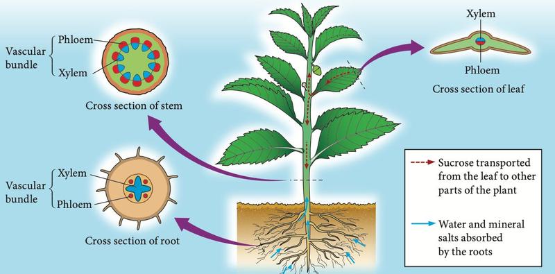
| Xylem | Phloem | |
|---|---|---|
| Transports | Water and dissolved mineral salts | Sucrose (food made in the leaves) |
| Direction | Roots to stem to leaves (upwards) | From the leaves to all other parts |
| Purpose | For photosynthesis and to replace water lost by transpiration | Food for growth, respiration and storage |
| Position in leaf | Upper side of the vascular bundle | Lower side of the vascular bundle |
Food: a ring of bark including phloem is removed from a woody branch. Sucrose cannot pass the gap, so it builds up and the bark swells above the ring; the plant below the ring is starved.
3.5Animals and plants compared
| Blood circulatory system (animals) | Transport system in plants | |
|---|---|---|
| Similarities | Both are transport systems; both carry water, nutrients and dissolved substances; both are found in complex organisms | |
| Structure | Tubular system with a heart (pump) and valves | Vessels without a pump or valves |
| Types of vessels | Three: artery, capillary, vein | Two: xylem and phloem |
| Connection | Arteries, capillaries and veins connect to form one continuous system | Xylem and phloem are separate and not connected |
Summary
| Remember | Key fact |
|---|---|
| Simple vs complex organisms | Simple: diffusion through the cell membrane. Complex: specialised transport system |
| Heart | 2 atria + 2 ventricles; left ventricle has the thickest wall; valves ensure one-way flow |
| Vessels | Artery: thick wall, pulse. Capillary: one cell thick, exchange. Vein: valves, large lumen |
| Double circulation | Pulmonary (heart, lungs, heart) and systemic (heart, body, heart) |
| Systole / diastole | Ventricles contract: "lub", higher pressure. Ventricles relax: "dub", lower pressure |
| Blood | Plasma 55%, red blood cells 45%, white blood cells and platelets under 1% |
| Groups | O = universal donor; AB = universal recipient |
| Transpiration | Water vapour lost mainly through stomata. Faster with more light, heat and wind; slower with high humidity |
| Xylem / phloem | Xylem: water and mineral salts up. Phloem: sucrose from leaves to all parts |