Homeostasis and the Human Urinary System
What this chapter covers
- Explain homeostasis and why physical and chemical factors of the internal environment must be kept within normal ranges (13.1.1, 13.1.2)
- Name the organ systems that regulate body temperature, blood sugar, partial pressure of carbon dioxide and blood pressure (13.1.3)
- Explain how negative feedback regulates each of these four factors (13.1.4)
- Identify the structure and functions of the kidney and the nephron (13.2.1, 13.2.2)
- Describe urine formation by ultrafiltration, reabsorption and secretion (13.2.3)
- Explain osmoregulation of blood osmotic pressure by ADH (13.2.4)
- Plan and interpret an experiment on water intake and urine volume (13.2.5)
- Describe kidney failure and kidney stones (13.3.1)
13.1Homeostasis
| Type | Factors regulated |
|---|---|
| Physical | Temperature, blood osmotic pressure, blood pressure |
| Chemical | pH, concentration of minerals, blood sugar |
Any deviation from the normal range triggers the homeostatic mechanism, which works by negative feedback: a value that is too high is brought down, and a value that is too low is raised, so the factor returns to the normal range.
| Factor | Organ systems involved |
|---|---|
| Body temperature | Integumentary (skin, sweat glands), nervous, circulatory, muscular, endocrine |
| Blood sugar level | Endocrine, circulatory, digestive |
| Partial pressure of CO2 | Respiratory, circulatory, nervous |
| Blood pressure | Circulatory, nervous |
Body temperature
Enzymes need an optimum temperature: too hot denatures them, too cold slows metabolism. Thermoreceptors in the skin and the hypothalamus (the temperature control centre) detect the change.
| Effector | Temperature rises | Temperature falls |
|---|---|---|
| Arterioles in skin | Vasodilation, more heat lost by radiation | Vasoconstriction, less heat lost |
| Sweat glands | More sweat; evaporation cools the skin | No sweating |
| Erector muscles and hair | Relax; hair lies flat, heat escapes | Contract; hair stands erect, trapped air insulates |
| Skeletal muscles | Do not shiver | Shiver, generating heat |
| Adrenal and thyroid glands | Less adrenaline and thyroxine; metabolic rate falls | More adrenaline and thyroxine; metabolic rate rises, more heat |
Blood sugar level
The normal range is 75–110 mg per 100 ml. The pancreas (islets of Langerhans) secretes the hormones that control it.
| After a meal: sugar rises | Between meals: sugar falls | |
|---|---|---|
| Cells stimulated | Beta cells | Alpha cells |
| Hormone | Insulin | Glucagon |
| Effect | Muscles use more glucose in respiration; excess glucose becomes glycogen in liver and muscle, and fat in adipose cells | Liver converts glycogen to glucose; fat is broken down to fatty acids for energy |
Partial pressure of carbon dioxide
During vigorous activity respiration raises CO2, which forms carbonic acid, so blood pH falls. Chemoreceptors (central, in the medulla oblongata; peripheral, in the carotid and aortic bodies) detect this. The respiratory control centre sends impulses so that the intercostal muscles, diaphragm and heart work faster. Breathing and heart rate rise, more CO2 is expelled, and CO2 pressure and pH return to normal.
Blood pressure
Baroreceptors in the aortic arch and carotid artery send impulses to the cardiovascular control centre in the medulla oblongata.
| Blood pressure rises | Blood pressure drops |
|---|---|
| Baroreceptors stimulated more; vasodilation; weaker heart contractions; pressure falls to normal | Baroreceptors stimulated less; vasoconstriction; stronger heart contractions; pressure rises to normal |
Show answerHide answer
13.2The human urinary system
The system has two kidneys, two ureters, the bladder and the urethra. The renal artery brings blood from the heart; the renal vein returns it. Urine flows from the kidney (pelvis) through the ureter to the bladder, then out through the urethra. Each kidney has a cortex and a medulla.
| Function | What the kidneys do |
|---|---|
| Excretion | Remove nitrogenous wastes: urea, uric acid, ammonia, creatinine |
| Osmoregulation | Control the volume of body water, ion concentration, blood osmotic pressure, electrolytes and pH |
The nephron and urine formation
The nephron is the functional unit of the kidney: Bowman's capsule with the glomerulus, then the proximal convoluted tubule, loop of Henle and distal convoluted tubule. Several nephrons join a collecting duct.
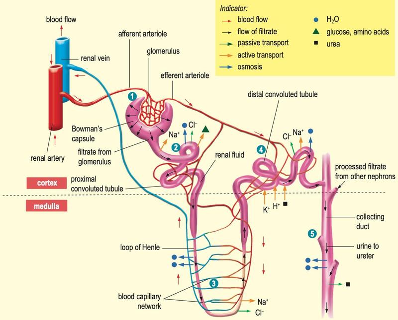
Figure: Biology Form 4 (DLP) textbook, p. 254.
| Stage | Where | What happens |
|---|---|---|
| 1 Ultrafiltration | Glomerulus to Bowman's capsule | The afferent arteriole is wider than the efferent, so pressure is high. Plasma without blood cells and proteins is forced out as glomerular filtrate. |
| 2 Reabsorption | Proximal convoluted tubule | Na+ actively pumped back, Cl– passive; all glucose and amino acids reabsorbed by active transport; water follows by osmosis. |
| 3 Reabsorption | Loop of Henle, distal tubule | Water by osmosis, Na+ by active transport. Amount depends on body water and salt. |
| 4 Secretion | Distal tubule and collecting duct | H+, K+, NH4+, urea, creatinine, toxins and drugs pass into the tubule by diffusion and active transport. |
| 5 Urine | Collecting duct | Remaining fluid is urine: water, urea, NaCl, uric acid, creatinine. |
Osmoregulation and ADH
| Too little water | Too much water | |
|---|---|---|
| Blood osmotic pressure | Rises | Falls |
| Osmoreceptors in hypothalamus | Stimulated | Less stimulated |
| ADH from pituitary | More | Less |
| Distal tubule and collecting duct | More permeable to water | Less permeable |
| Urine | Small volume, concentrated | Large volume, dilute |
13.3Health issues of the urinary system
- Kidney failure can follow illness, bacterial infection or accident. Diabetes mellitus is the leading cause, then high blood pressure; both damage the glomerulus. Patients may need haemodialysis.
- Kidney stones are hard masses of uric acid, calcium oxalate or calcium phosphate. They can block the ureter and reduce urine production. Drinking plenty of water lowers the risk.