Biology notes, chapter 14
Support and Movement in Humans and Animals
Textbook Biology Form 4 KSSM (DLP), pages 264–283
What this chapter covers
- List the types of skeletons in humans and animals and justify why skeletons are needed (14.1)
- Identify the bones of the axial and appendicular skeletons (14.2.1)
- Characterise and compare the cervical, thoracic, lumbar, sacral and caudal vertebrae (14.2.2, 14.2.3)
- State the types of joints and draw, label and explain the structure of a hinge joint (14.2.4, 14.2.5)
- Explain the movement of the human forearm and leg, and the locomotion of fish, birds, earthworms and grasshoppers (14.3)
- Describe health issues of the musculoskeletal system and justify practices to keep it healthy (14.4)
14.1Types of skeletons
A skeleton supports the body, protects soft organs and lets an organism move from place to place. There are three types: hydrostatic skeleton, exoskeleton and endoskeleton.
| Type | Description | Example |
|---|---|---|
| Hydrostatic skeleton | Fluid held under pressure in a body cavity, surrounded by muscles. Keeps a soft body in shape. | Earthworm |
| Exoskeleton | Hard covering on the outside of the body. Supports and protects organs; muscles attach to its inner surface. | Grasshopper, crab |
| Endoskeleton | Skeleton of bone or cartilage inside the body. Keeps the body shape, supports soft tissues and protects internal organs. | Humans, fish, birds |
Why skeletons are needed
- Support: soft body tissues cannot hold the body shape on their own.
- Protection of internal organs from injury (for example the rib cage protects the heart and lungs).
- Movement: muscles pull on skeletal parts so the organism can look for food and partners or escape from predators.
Why does an earthworm need a hydrostatic skeleton?
Show answerHide answer
Its body is soft; the fluid under pressure keeps its body rigid and in shape so that muscles can work against it.
14.2Musculoskeletal system of humans
Axial and appendicular skeletons
| Part | Bones |
|---|---|
| Axial skeleton | Skull, vertebral column, ribs, sternum (breastbone) |
| Appendicular skeleton | Pectoral girdle (clavicle, scapula), pelvic girdle (ilium, pubis, ischium), upper limbs, lower limbs |
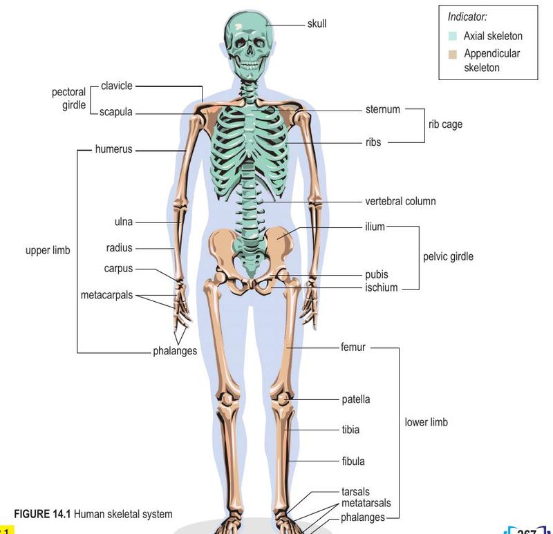
Upper limb: humerus, radius, ulna, carpals, metacarpals, phalanges. Lower limb: femur, patella, tibia, fibula, tarsals, metatarsals, phalanges.
Vertebrae
Every vertebra has a neural canal that holds the spinal cord, a spinous process and transverse processes for muscle and ligament attachment, and a centrum (except the atlas).
| Vertebra | Characteristics |
|---|---|
| Cervical | Short spinous process; wide, short transverse processes; small centrum; a pair of transverse foramina |
| Atlas (1st cervical) | Large neural canal, small spinous process, no centrum; articulates with the skull |
| Axis (2nd cervical) | Large spinous process, small transverse processes; odontoid process fits into the atlas |
| Thoracic | Long spinous and transverse processes; medium centrum; facets for ribs; no transverse foramen |
| Lumbar | Biggest and strongest; short spinous and transverse processes; large centrum to bear the weight of the lower back |
| Sacral | Five vertebrae fused into a triangular sacrum; passage for nerves and blood vessels |
| Caudal | Four vertebrae fused into a small pointed structure |
Careful! Only cervical vertebrae have transverse foramina. Do not give them to thoracic or lumbar vertebrae.
Joints
| Type | Example |
|---|---|
| Immovable | Sutures in the cranium of the skull |
| Slightly moveable | Cartilage discs between vertebrae; cartilage between first rib and sternum |
| Freely moveable | Hinge joint (elbow, knee, fingers); ball-and-socket joint (shoulder, hip) |
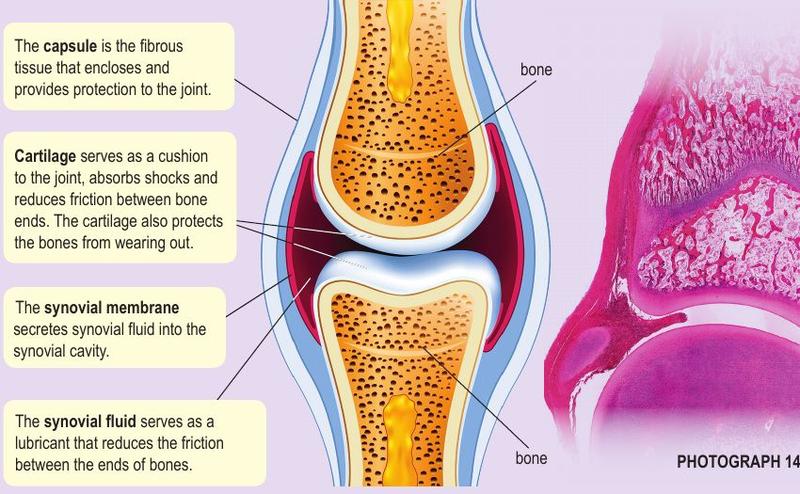
A hinge joint moves in one plane only (back and forth). A ball-and-socket joint rotates in all directions.
| Part | Function |
|---|---|
| Cartilage | Cushion; absorbs shock; reduces friction; stops bone ends wearing out |
| Synovial membrane | Secretes synovial fluid |
| Synovial fluid | Lubricant that reduces friction between bone ends |
| Capsule | Fibrous tissue that encloses and protects the joint |
| Skeletal muscle | Bundles of muscle fibres with many nerves and blood vessels; works in antagonistic pairs |
| Tendon | Joins muscle to bone; strong, flexible but not elastic |
| Ligament | Joins bone to bone; strong, tough and elastic; supports the joint |
Memory aid: Tendon joins Two different things (muscle to bone). Ligament joins Like with like (bone to bone).
State two differences between a tendon and a ligament.
Show answerHide answer
A tendon connects muscle to bone and is not elastic; a ligament connects bone to bone and is elastic.
14.3Movement and locomotion
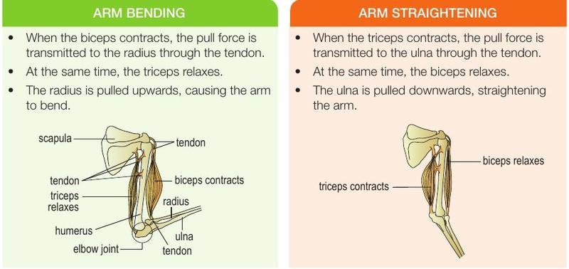
Antagonistic muscles work in pairs in opposite directions: when one contracts the other relaxes. A flexor bends a limb; an extensor straightens it.
Human forearm
| Bending the arm | Straightening the arm | |
|---|---|---|
| Biceps | Contracts | Relaxes |
| Triceps | Relaxes | Contracts |
| Bone pulled | Radius is pulled upwards (through the tendon) | Ulna is pulled downwards (through the tendon) |
Human leg (walking)
The biceps femoris (flexor) contracts to bend the leg at the knee and lift it. The quadriceps femoris (extensor) contracts to straighten the leg. The calf muscle lifts the heel and the tibialis brings the heel down. The sequence then repeats with the other leg.
Locomotion in animals
| Animal | Mechanism |
|---|---|
| Fish | Myotomes (W-shaped muscle segments) on the two sides contract and relax alternately, whipping the tail from side to side and pushing water backwards and sideways. Fins control direction. |
| Bird | Pectoralis major contracts (minor relaxes) to pull the wings down; pectoralis minor contracts (major relaxes) to pull them up. |
| Earthworm | Circular and longitudinal muscles contract alternately in a wave of peristalsis. Chaetae anchor segments to the ground. |
| Grasshopper | Flexor contracts to fold the hind leg into a Z shape; extensor contracts to straighten it suddenly and launch the insect forward and up. |
Worked example: earthworm
When the longitudinal muscles contract and circular muscles relax, the earthworm becomes shorter and thicker. When the circular muscles contract and longitudinal muscles relax, it becomes longer and thinner and the front end extends forward.
What is the role of the myotomes in a swimming fish?
Show answerHide answer
They are antagonistic muscle segments; alternate contraction and relaxation on each side whips the tail and pushes the fish forward.
14.4Health issues of the musculoskeletal system
| Disorder | Cause | Effect |
|---|---|---|
| Osteoporosis | Calcium loss faster than absorption with age; lack of exercise; low calcium, phosphorus and vitamin D; low oestrogen after menopause | Thin, porous, brittle bones; reduced height; stooped posture |
| Osteomalacia | Lack of calcium, phosphorus and vitamin D (adults, especially pregnant women) | Soft bones |
| Rickets | Same deficiency in children | Soft, weak bones with defects |
| Arthritis (osteoarthritis) | Less synovial fluid; wear and tear of cartilage | Swollen, painful, stiff joints |
| Scoliosis | Genetic factor or abnormal growth of the backbone during puberty | Backbone bent sideways in an S or C shape |
Keeping the system healthy
- Good posture: slouching strains the vertebral column.
- Proper attire: loose clothes and low-heeled cushioned shoes.
- Exercise: strengthens joints, bones and muscles; helps prevent osteoporosis.
- Balanced diet: calcium, phosphate, vitamin C and D. Vitamin D helps calcium absorption.
Why is osteoporosis common in women after menopause?
Show answerHide answer
Oestrogen level falls. Oestrogen helps calcium absorption and reduces calcium loss from bones, so bone density drops.