Sexual Reproduction, Development and Growth in Humans and Animals
What this chapter covers
- Describe the structure and functions of the male and female reproductive systems (15.1)
- Explain the necessity of gametogenesis; describe spermatogenesis and oogenesis and compare them (15.2)
- Analyse hormone levels, follicle growth and endometrial changes in the menstrual cycle, and in pregnancy and miscarriage (15.3)
- Describe fertilisation, early embryo development, the role of HCG, the placenta and the umbilical cord (15.4)
- Describe the formation of identical, fraternal and conjoined twins (15.5)
- State the causes of impotency and its treatments (15.6)
- Explain growth, how it is measured, metamorphosis, sigmoid and intermittent growth curves (15.7)
15.1Reproductive system of humans
| Organ | Function |
|---|---|
| Testis | Produces sperm and the male sex hormone testosterone |
| Scrotum | A sac that holds and protects the testes |
| Sperm duct (vas deferens) | Carries sperm |
| Seminal vesicle | Secretes a fluid with nutrients for the sperm |
| Prostate gland | Secretes a fluid that helps sperm movement |
| Urethra | Tube that carries sperm and urine out of the body |
| Penis | Releases sperm into the vagina during copulation |
| Ovary | Produces ova and the hormones oestrogen and progesterone |
| Fallopian tube | Lined with cilia; cilia and peristalsis move the secondary oocyte or embryo to the uterus. Fertilisation happens here. |
| Uterus | Thick muscular wall; endometrium receives and nourishes the embryo |
| Cervix | Narrow opening of the uterus; mucus helps sperm swim up |
| Vagina | Where sperm enter; passage for birth and menstruation |
15.2Gametogenesis in humans
Spermatogenesis (seminiferous tubules of the testes)
Primordial germ cell → (mitosis) spermatogonium 2n → primary spermatocyte 2n (growth) → (meiosis I) two secondary spermatocytes n → (meiosis II) four spermatids n → (differentiation) four sperms. FSH and testosterone stimulate it; LH stimulates testosterone secretion. Sertoli cells nourish the developing cells.
Oogenesis (ovaries)
- Before birth: oogonium (2n) forms a primary oocyte, which begins meiosis I and stops at prophase I.
- From puberty, each month: one primary oocyte completes meiosis I, giving a secondary oocyte and a first polar body. The secondary oocyte stops at metaphase II.
- Ovulation: the Graafian follicle releases the secondary oocyte. Meiosis II is completed only if a sperm penetrates it, giving the ovum and a polar body.
- The empty follicle becomes the corpus luteum.
Structure of the gametes
- Sperm: head (nucleus), midpiece (many mitochondria for energy) and tail (swimming).
- Graafian follicle: a large secondary oocyte surrounded by a gel-like layer and follicular cells.
| Feature | Spermatogenesis | Oogenesis |
|---|---|---|
| Site | Testes | Ovaries |
| Gametes from one germ cell | Four sperms | One secondary oocyte and polar bodies that die |
| Meiosis | Completed | Meiosis II completed only after fertilisation |
| Start | Puberty, continuous to old age | Before birth; paused; resumes at puberty; stops at menopause |
| Number | Millions every day | One per menstrual cycle |
Show answerHide answer
15.3Menstrual cycle
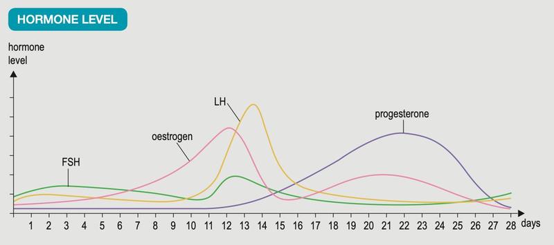
Textbook Figure 15.9, p. 294 (hormone graph).
| Hormone | Source | Function |
|---|---|---|
| FSH | Pituitary | Stimulates follicle growth and oestrogen release |
| LH | Pituitary | Stimulates ovulation, corpus luteum formation and progesterone release |
| Oestrogen | Ovary (follicle, corpus luteum) | Repairs the endometrium; matures the follicle; peak triggers the LH surge (positive feedback) |
| Progesterone | Corpus luteum | Thickens the endometrium; inhibits FSH and LH (negative feedback) |
| Days | Events |
|---|---|
| 0–5 | Endometrium sheds (menstruation). GnRH makes the pituitary release FSH and LH; follicles grow. |
| 6–14 | Oestrogen peaks on day 12; LH peaks on day 13; ovulation on day 14. The follicle becomes the corpus luteum. |
| 15–21 | Corpus luteum secretes progesterone and oestrogen; endometrium thickens; FSH and LH are inhibited. |
| 22–28 | No fertilisation: corpus luteum degenerates, hormones fall, endometrium sheds. |
Pregnancy, miscarriage and syndromes
- If fertilisation occurs, the corpus luteum keeps secreting progesterone and oestrogen for three to four months, then the placenta takes over until birth. Progesterone prevents FSH and LH release, so there is no ovulation or menstruation in pregnancy.
- A fall in progesterone can cause miscarriage.
- Premenstrual syndrome: symptoms 7 to 14 days before menstruation, caused by oestrogen and progesterone imbalance.
- Menopause: about 46 to 50 years, ovulation and menstruation stop because oestrogen and progesterone fall.
Show answerHide answer
15.4Development of a human foetus
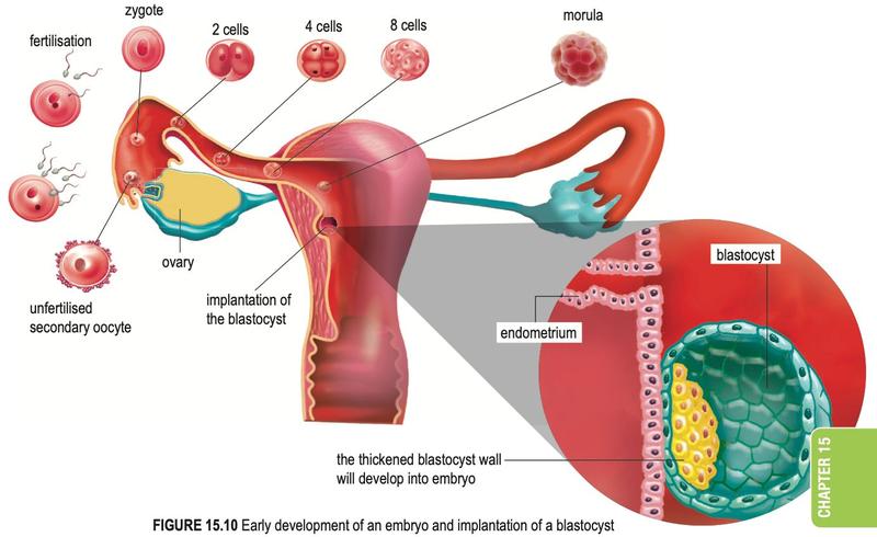
Textbook Figure 15.10, p. 297.
- One sperm penetrates the secondary oocyte in the Fallopian tube; the membrane changes so no other sperm enters. The nuclei fuse to form a diploid zygote.
- The zygote divides by mitosis: two-cell embryo, morula, then blastocyst.
- The blastocyst implants in the endometrium.
Placenta and umbilical cord
- Umbilical vein: oxygenated blood with nutrients, placenta to foetus.
- Umbilical arteries: deoxygenated blood with carbon dioxide and urea, foetus to placenta.
- Placenta: exchange of glucose, amino acids, antibodies, oxygen in and carbon dioxide, urea out; also secretes progesterone and oestrogen from month 4.
Why foetal and maternal blood do not mix (a thin membrane separates them): it protects the foetus from toxins and bacteria; prevents foetal vessels bursting under the mother's higher blood pressure; prevents agglutination if blood groups differ. Drugs, alcohol, nicotine and viruses such as HIV and rubella can still cross.
15.5Formation of twins
| Feature | Identical twins | Fraternal twins |
|---|---|---|
| Origin | One ovum, one sperm: one zygote that divides into two embryos | Two ova, two sperms: two zygotes |
| Placenta | One shared | Separate |
| Genes | Same | Different |
| Sex | Always the same | Same or different |
Conjoined twins arise when the embryo of identical twins divides incompletely, so parts such as the chest or abdomen stay fused. Separation by surgery depends on the organs they share.
15.6Impotency
Impotency means a couple cannot conceive. Male causes: erectile dysfunction, testes not producing sperm, low or abnormal sperm, hormonal imbalance. Female causes: ovaries not producing secondary oocytes, blocked Fallopian tubes, abnormal uterus, hormonal imbalance. Treatments: hormone therapy; surgery for blocked tubes or ducts; in vitro fertilisation (IVF) for blocked Fallopian tubes.
15.7Growth in humans and animals
- Parameters: height or length; fresh mass (organism survives, less accurate because of water); dry mass (dried at 100 °C until constant, organism is killed, more accurate).
- Complete metamorphosis (butterfly): egg, larva, pupa, adult.
- Incomplete metamorphosis (grasshopper): egg, nymph (several ecdyses), adult. No pupa.
| Phase of sigmoid curve | Growth rate | Key events |
|---|---|---|
| Lag | Low | Adjusting to environment; little cell division |
| Exponential | Highest | Active cell division and elongation |
| Stationary | Slows, constant | Cells reach maximum size, differentiation |
| Maturity | Zero | Cell division only replaces dead cells |
| Senescence | Negative | Ageing |
| Death | — | Ageing, illness or lack of nutrients |
Intermittent growth in insects: the hard exoskeleton cannot expand, so the insect moults (ecdysis, controlled by hormones). The graph is a staircase: flat parts are instars (no length increase), steep parts are ecdysis with rapid growth.